- 9 de Junho de 2026 - Terça
TUBERCULOSE ENDOBRÔNQUICA
Tuberculose endobrônquicaPESQUISA DE IMAGENS
- ABSCESSO PULMONAR
- ANATOMIA RADIOLÓGICA
- ASMA
- BRONQUIECTASIAS
- BRONQUIOLITES
- DIVERSOS
- DOENÇA DO REFLUXO GASTRESOFÁGICO
- DOENÇAS FÚNGICAS
- DOENÇAS INTERSTICIAIS
- DOENÇAS OCUPACIONAIS
- DOENÇAS PLEURAIS
- DOENÇAS VASCULARES
- DPOC
- METÁSTASES
- MICOBACTERIOSES NÃO-TUBERCULOSAS
- NEOPLASIAS
- NÓDULOS
- OUTROS ÓRGÃOS
- PNEUMONIAS
- TUBERCULOSE
- VIAS AÉREAS SUPERIORES
TUBERCULOSE ENDOBRÔNQUICA

Tuberculose endobrônquica em brônquio fonte esquerdo ocasionando atelectasia parcial de lobo superior esquerdo (seta amarela).
Chaves: tbc.
*****
Endobronchial tuberculosis in main left bronchus causing partial atelectasis of the left upper lobe (yellow arrow).
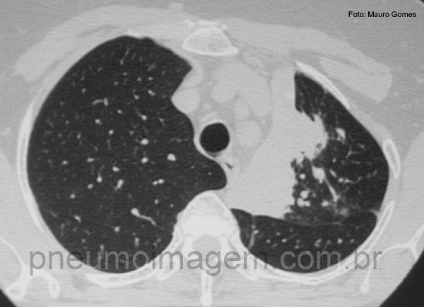
Neste corte tomográfico se observa a atelectasia parcial do lobo superior esquerdo. Há nódulos centrolobulares na periferia da lesão que podem corresponder a disseminação broncogênica da tuberculose.
*****
In this CT slice is observed partial atelectasis of the left upper lobe. There are centrilobular nodules in the periphery of the lesion which may correspond to bronchogenic spread of tuberculosis.
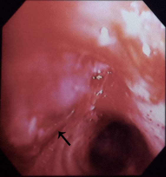
Nesta imagem obtida por broncoscopia de outro caso semelhante, observa-se oclusão do brônquio do lobo superior por lesão de aspecto liso e violáceo (seta). O anatomopatológico mostrou processo inflamatório crônico granulomatoso com necrose caseosa e pesquisa de BAAR positiva no material.
*****
In this image obtained by bronchoscopy from another patient, there is occlusion of the upper lobe bronchus, which is filled by a smooth and violaceous lesion (arrow). Histopathological examination revealed chronic granulomatous inflammation with caseous necrosis and AFB smear positive.


DEIXE SEU COMENTÁRIO