- 24 de Julho de 2026 - Sexta
TROMBOEMBOLIA PULMONAR
Tromboembolia pulmonarPESQUISA DE IMAGENS
- ABSCESSO PULMONAR
- ANATOMIA RADIOLÓGICA
- ASMA
- BRONQUIECTASIAS
- BRONQUIOLITES
- DIVERSOS
- DOENÇA DO REFLUXO GASTRESOFÁGICO
- DOENÇAS FÚNGICAS
- DOENÇAS INTERSTICIAIS
- DOENÇAS OCUPACIONAIS
- DOENÇAS PLEURAIS
- DOENÇAS VASCULARES
- DPOC
- METÁSTASES
- MICOBACTERIOSES NÃO-TUBERCULOSAS
- NEOPLASIAS
- NÓDULOS
- OUTROS ÓRGÃOS
- PNEUMONIAS
- TUBERCULOSE
- VIAS AÉREAS SUPERIORES
TROMBOEMBOLIA PULMONAR

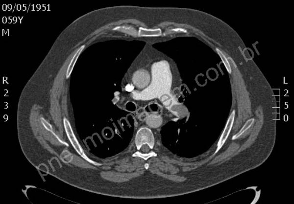
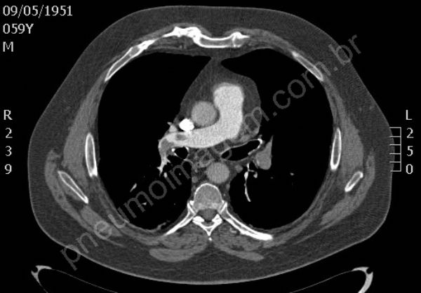
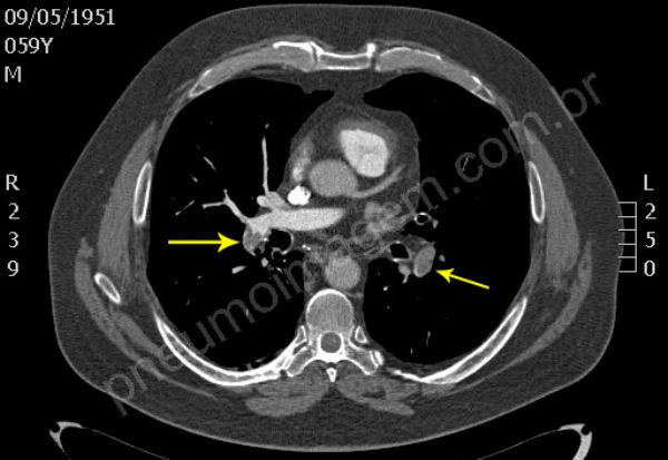
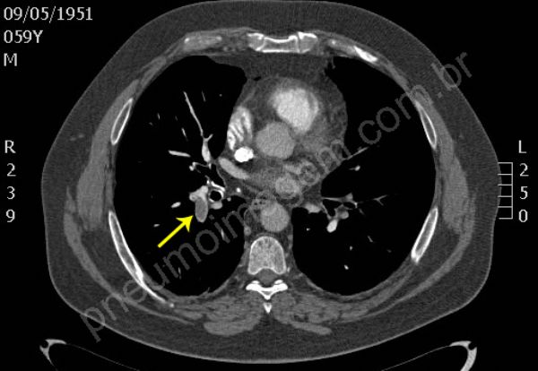
Tromboembolia pulmonar (TEP). Na sequência tomográfica observam-se trombos localizados no tronco da artéria pulmonar e nos principais ramos segmentares à esquerda. Os trombos na bifurcação do tronco pulmonar à direita estendem-se para ramos segmentares do lobo superior, para a interlobar descendente e ramos segmentares do lobo médio. O paciente apresentava também trombose venosa profunda (TVP) na veia femural esquerda, fator de risco importante para TEP. Chaves: tromboembolismo, embolia pulmonar.
*****
Pulmonary thromboembolism (PTE). In tomographic sequence are observed thrombi located in the main pulmonary artery and major segmentary branches left. Thrombi in the bifurcation of the pulmonary trunk right extend to segmentary branches of the upper lobe, extend to descending interlobar and segmentary branches of the middle lobe. The patient also presented deep vein thrombosis (DVT) in the left femoral vein, an important risk factor for PE. Keys: pulmonary embolism (PE).
Exames de imagem possuem um papel essencial no diagnóstico de embolia pulmonar (TEP). Estudos demonstraram que o TEP pode ser afastado em pacientes com baixa probabilidade clínica e dímero-D normal. Para todos os outros, exames de imagem são necessários e a angiotomografia computadorizada é o exame de escolha devido à sua alta sensibilidade e especificidade. Cintilografia de ventilação e perfusão é reservada para pacientes com contra-indicação para a TC (Semin Respir Crit Care Med. 2012;33(2):138-143).
*****
Imaging tests have an essential role in the diagnosis of pulmonary embolism (PE). Studies have shown that diagnosis of PTE may be removed in patients with low clinical probability and D-dimer normal. For everyone else, imaging tests are needed and CT angiography is the method of choice because of its high sensitivity and specificity. Ventilation and perfusion scintigraphy is reserved for patients with contraindications for TC (Semin Respir Crit Care Med 2012; 33 (2):. 138-143).


DEIXE SEU COMENTÁRIO